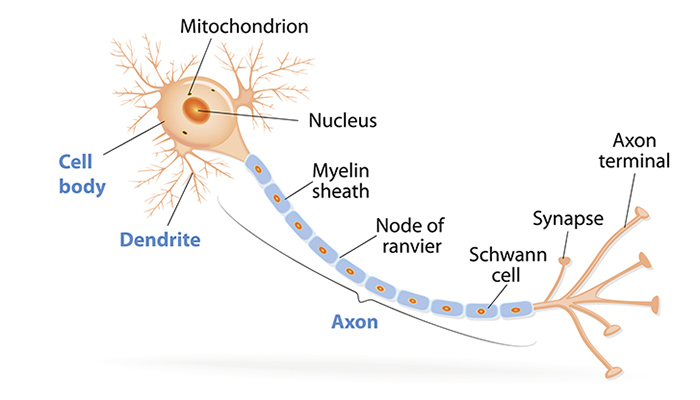
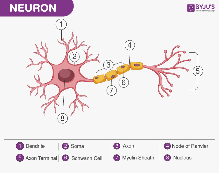
10+ label the structures of the neuron in the diagram
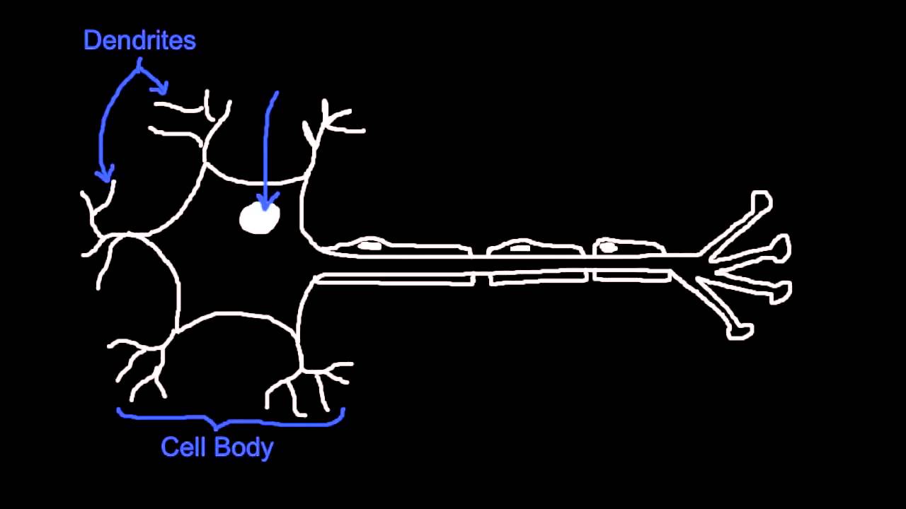
What Does a Neuron Look Like The main. A region for receiving and processing incoming information from other cells.
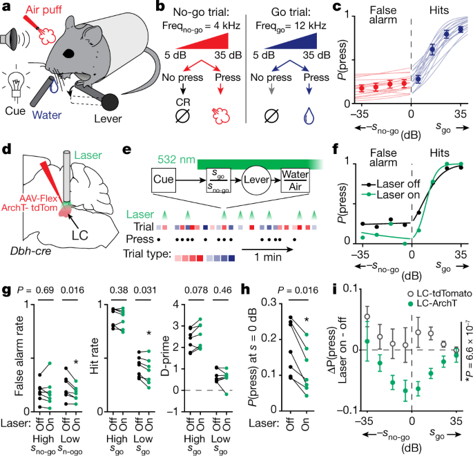
Spatiotemporal Dynamics Of Noradrenaline During Learned Behaviour Nature
View Anatomy of a Neurondocx from ANAT PHYSIO HON P33P33 at South Broward High School.
. Learn vocabulary terms and more with flashcards games and other study tools. The message then moves through the axon to the other end of the neuron then to the tips of the axon and then into the space between neurons. Each neuron has a cell body which is the central area of the neuron.
The neuron is broken up into two major regions. These are branch-like structures that receive messages from other neurons and allow the transmission of messages to the. A neuron is a structural and functional unit of the neural tissue and hence the neural system.
Terminal buttons dendrites myelin sheath cell body axon synapse Drag each item above to its appropriate location in the image. Start studying Label the Structures of the Neuron. Although neurons look complicated their design is actually quite simple.
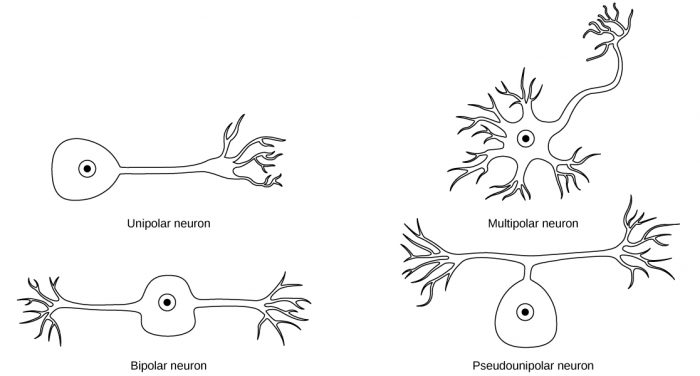
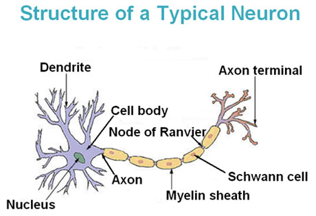
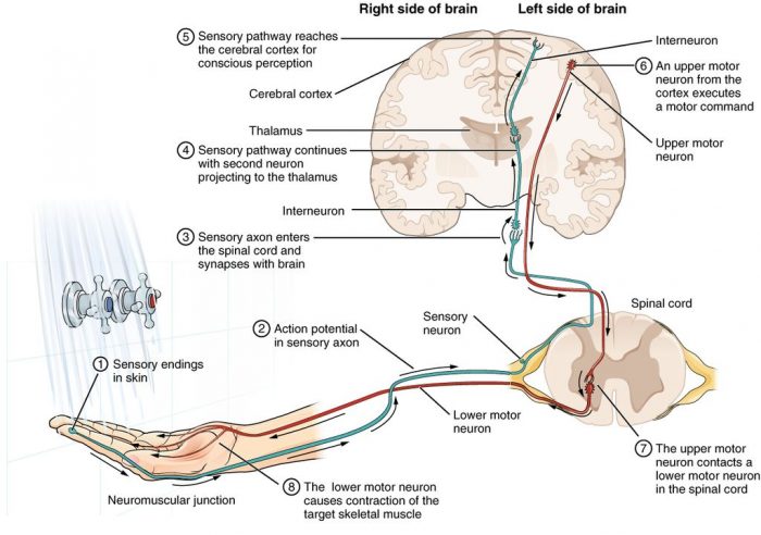
Label the following structures on the diagram of a multipolar neuron shown below. Neurons pass messages to each other using a special type of electrical signal. The Structure of a Neuron.
Creating a neuron-labeled image by hand can be difficult. The cell bodythe part of the cell that houses the nucleus and keeps the entire cell alive and functioning. Dendrites are extensions of the cell body or soma of a neuron that receive and convey signals back to the cell body.
Thus neurons with longer processes projections are the longest cells in the body. Structure of Neuron. Human neural system has about 100 billion neurons.
Dendrites receive messages from other neurons. Certain neurons may almost equal the length of body itself. Cell body nucleus nucleolus chromatophilic substance dendrites initial segment of axon myelin sheath.
Anatomy of a Neuron Student. The cell body expands into an axon which is encased in. The type of information that is received processed.
Following are the different parts of a neuron. Complete the charts and label the. Receives impulses from other nerve cells.
It contains the nucleus and other structures common to all cells in the body such as. All neurons have three different parts dendrites cell body and axon. Majority of the neurons occur in the brain.
From there the message can move to the next neuron. A neuron varies in shape and size depending on its function and location. Label the structures of the neuron in the diagram.
The students must use the EdrawMax Online tool to make a high-quality neuron diagram. A region for conducting and transmitting information to other cells.
A Labelled Diagram Of Neuron With Detailed Explanations

Labeled Neuron Diagram Science Trends

A P2 Lab 1 Hw Flashcards Quizlet
Labeled Neuron Diagram Science Trends

Act 5 Human Body Docx Part 1 1 Build A Clay Model Of A Motor Neuron Using A Different Color Clay For The Different Parts Of The Neuron And Then Course Hero

Label The Structures Of The Neuron Diagram Quizlet
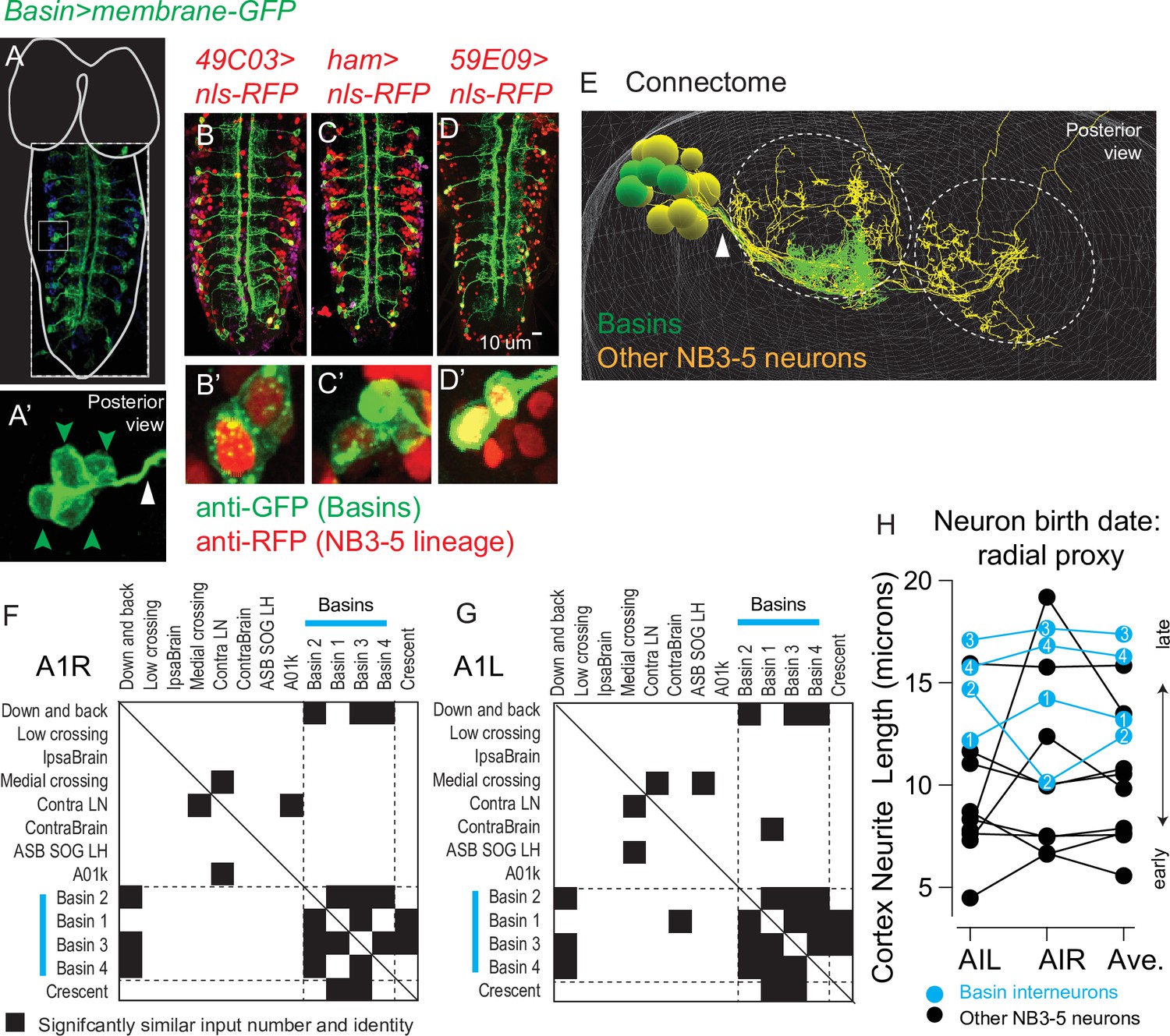
Sequential Addition Of Neuronal Stem Cell Temporal Cohorts Generates A Feed Forward Circuit In The Drosophila Larval Nerve Cord Elife

Label The Parts Of A Neuron Neurons Teaching Biology Technology Life
Neuron Creations Wikieducator
Solved Label The Parts Please Course Hero

Cell Nucleus Wikiwand
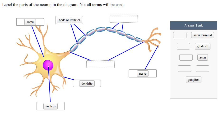
Solved Label The Parts Of The Neuron In The Diagram Not All Chegg Com
6 5 2 Draw And Label A Diagram Of The Structure Of A Motor Neuron Youtube
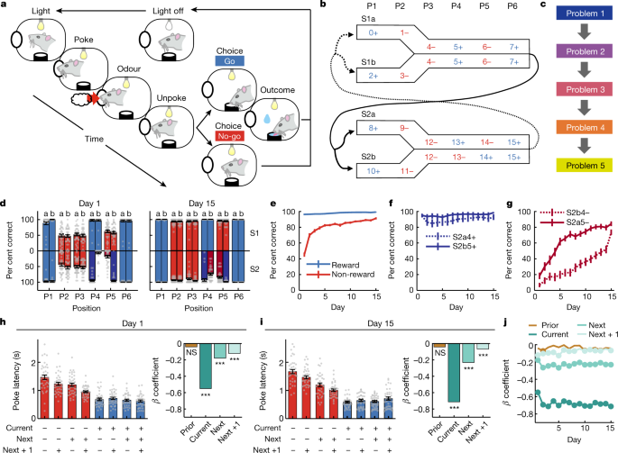
Evolving Schema Representations In Orbitofrontal Ensembles During Learning Nature
Neuroscience For Kids Baw Lesson Plan
Labeled Neuron Diagram Science Trends

Glia Actively Sculpt Sensory Neurons By Controlled Phagocytosis To Tune Animal Behavior Biorxiv